3D Brain Model
Turn this labeled brain around to see how its main regions fit together. The model shows the four lobes of the cerebrum, the deep structures in the middle, and the cerebellum and brainstem below, and each part is explained below.
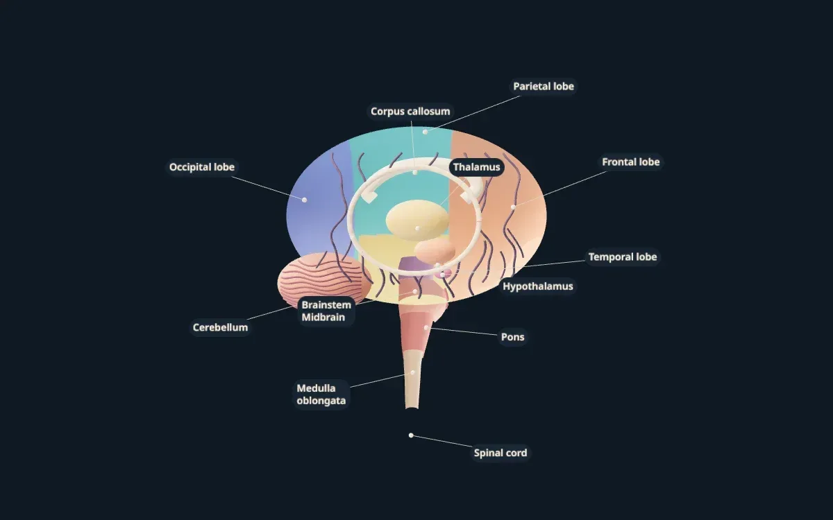
An illustrative teaching model built with Scholarly. Shapes, sizes, and colors are simplified so each part is easy to see.
Free to start. Build one from your own notes, slides, or textbook PDF.
Labeled parts
Parts of the brain
The 12 parts labeled on the model, and what each one does.
- Frontal lobe
- At the front of the brain. It handles planning, decisions, personality, speech production, and voluntary movement.
- Parietal lobe
- Behind the frontal lobe. It processes touch, temperature, and pain, and helps you sense where your body is in space.
- Temporal lobe
- On each side, near the ears. It processes hearing and helps with language and forming memories.
- Occipital lobe
- At the back of the brain. It is the main center for vision.
- Corpus callosum
- A thick band of nerve fibers that connects the left and right halves of the brain so they can share information.
- Thalamus
- A relay station deep in the brain that passes most sensory signals on to the right part of the cortex.
- Hypothalamus
- A small region below the thalamus that controls body temperature, hunger, thirst, sleep, and hormones through the pituitary gland.
- Cerebellum
- Under the back of the brain. It coordinates movement, balance, and posture.
- Midbrain
- The top part of the brainstem. It helps with eye movement and with reflexes to sight and sound.
- Pons
- The middle part of the brainstem. It relays signals between the cerebrum and cerebellum and helps control breathing.
- Medulla oblongata
- The lowest part of the brainstem. It controls automatic functions such as heartbeat, breathing, and blood pressure.
- Spinal cord
- Continues down from the brainstem and carries signals between the brain and the rest of the body.
For class and projects
Using this model for class or a project
For studying, rotate the model and name each lobe with one job it does: frontal for planning, parietal for touch, temporal for hearing, occipital for vision. Then work down the brainstem from the midbrain to the medulla.
For a brain model project, use it as a reference for proportions. The cerebrum is by far the largest part, the cerebellum tucks under the back, and the brainstem connects straight down into the spinal cord.
Teachers can show the model on a classroom screen while explaining how regions share work. For a psychology or neuroscience course that needs the limbic system, cranial nerves, or Brodmann areas, you can build your own version from your notes.
Make your own
Need a model of your own lesson?
This model was built in Scholarly from a short description. You can build one from your own material, labeled in the words your course uses.
- 01
Add your material or describe it
Pick notes, slides, a textbook PDF, or a lecture recording, or type what the model should show.
- 02
Scholarly builds the model
In a few minutes you get an interactive 3D model with labels. A full build uses 1 AI Creation Credit, and you confirm it first.
- 03
Refine it, then use it
Ask for small changes in chat for free. On a paid plan, export a 4K picture, a turntable video, or a .glb file for slides.
FAQ
Brain model questions
What are the main parts of the brain?
The cerebrum, divided into the frontal, parietal, temporal, and occipital lobes; the deep structures such as the thalamus and hypothalamus; the cerebellum; and the brainstem, made of the midbrain, pons, and medulla oblongata.
What are the four lobes of the brain?
The frontal lobe (planning, movement, speech), the parietal lobe (touch and body position), the temporal lobe (hearing and memory), and the occipital lobe (vision).
Is this brain model anatomically exact?
No. It is a simplified teaching model. Regions are colored and spaced so each one is easy to see. It is not a medical atlas and is not for clinical use.
Can I use this model for a school project?
Yes, you can use it as a reference while you study or build a physical model. Exporting pictures or video of models you build yourself needs a paid plan.
Can I make my own 3D brain model?
Yes. Sign up, choose 3D model, and describe what you need or add your notes. Scholarly builds a labeled, interactive model in a few minutes. A full build uses 1 AI Creation Credit, and you confirm it first.
Keep exploring
More interactive 3D models
Animal cell 3D model
A labeled cutaway with the nucleus, mitochondria, ER, and Golgi.
Plant cell 3D model
Cell wall, chloroplasts, and the large central vacuole, labeled.
Human heart 3D model
Four chambers, four valves, and the great vessels in a cutaway.
Human skull 3D model
The cranial and facial bones and the main sutures.
Human skeleton 3D model
The major bones of the body, labeled head to toe.
Human eye 3D model
Cornea, lens, retina, and optic nerve in a cutaway.
DNA 3D model
The double helix with its backbone and base pairs.
Atom 3D model
A carbon atom's nucleus and electron shells, Bohr style.
Solar system 3D model
The Sun and eight planets on their orbits.
3D models in Scholarly
How 3D models work, what they cost, and what they can't do yet.
Turn your notes into a 3D model
Build a labeled, interactive model of whatever you are studying or teaching, then make flashcards and a quiz from the same notes.
3D models are in Beta. A full build uses 1 AI Creation Credit; small chat edits are free.