Animal Cell 3D Model
Turn this labeled animal cell around to see how its organelles sit inside the cell membrane. The cutaway opens the cell so the nucleus, mitochondria, endoplasmic reticulum, and Golgi apparatus are all visible at once, and each part is explained below.
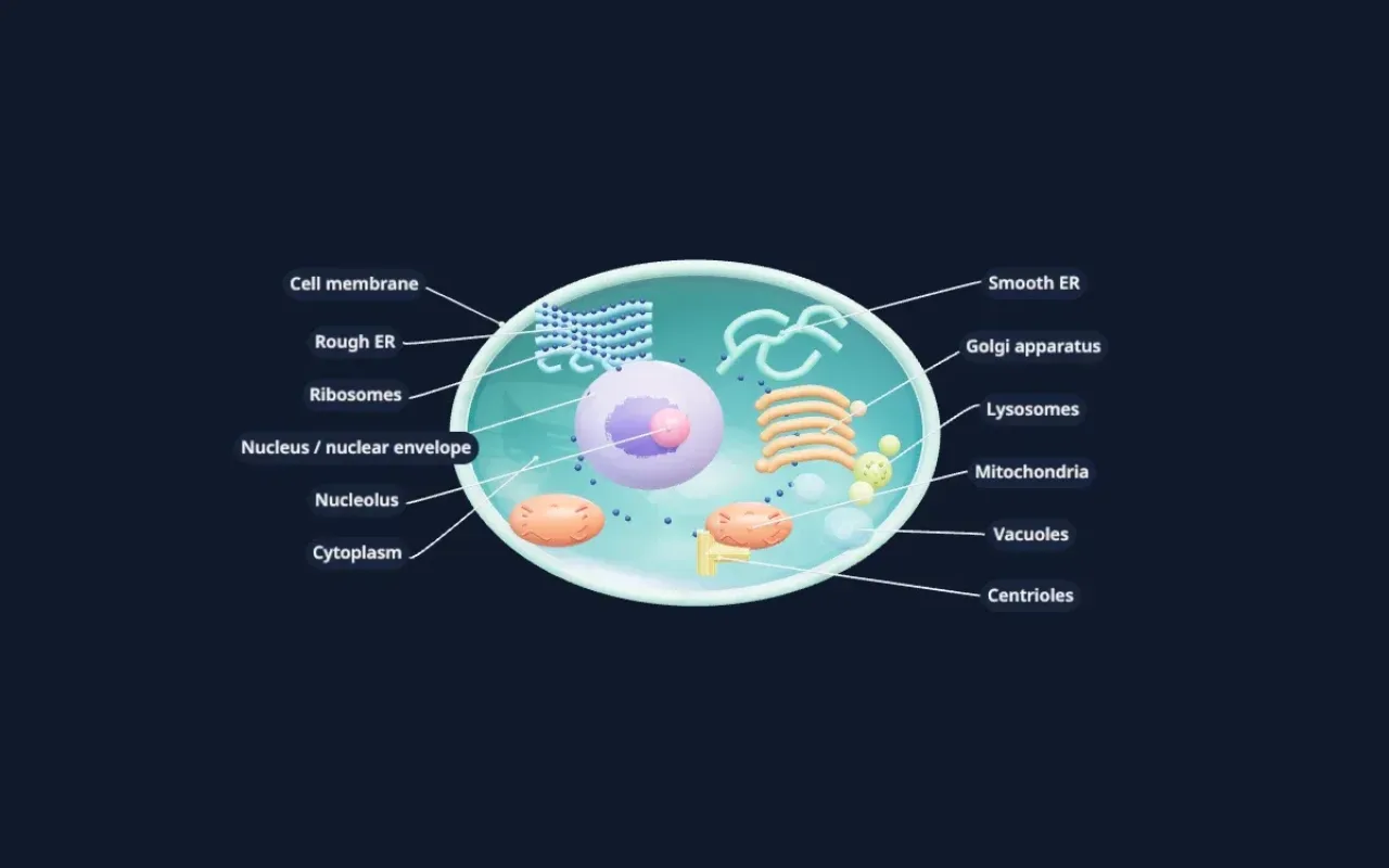
An illustrative teaching model built with Scholarly. Shapes, sizes, and colors are simplified so each part is easy to see.
Free to start. Build one from your own notes, slides, or textbook PDF.
Labeled parts
Parts of an animal cell
The 11 parts labeled on the model, and what each one does.
- Cell membrane
- A thin, flexible layer of lipids and proteins around the cell. It controls what enters and leaves.
- Cytoplasm
- The jelly-like fluid that fills the cell and holds the organelles in place. Many chemical reactions happen here.
- Nucleus and nuclear envelope
- The nucleus holds the cell's DNA and directs its activity. The nuclear envelope is the double membrane around it, with pores that let molecules in and out.
- Nucleolus
- A dense region inside the nucleus where ribosomes start to be made.
- Rough endoplasmic reticulum
- Folded membranes studded with ribosomes. Proteins made here are folded and sent on to the Golgi apparatus.
- Smooth endoplasmic reticulum
- Membrane tubes without ribosomes. They make lipids and help break down some toxins.
- Ribosomes
- Tiny structures that build proteins from the instructions in RNA. Some float free; others sit on the rough ER.
- Golgi apparatus
- A stack of flattened sacs that modifies, sorts, and packages proteins and lipids for use inside or outside the cell.
- Mitochondria
- The site of cellular respiration, which releases energy from food as ATP. Often called the powerhouse of the cell.
- Lysosomes
- Small sacs of digestive enzymes that break down waste, worn-out organelles, and things the cell takes in.
- Centrioles and vacuoles
- Centrioles are paired cylinders that help organize the cell during division. Animal cells have small vacuoles that store water and other substances.
For class and projects
Using this model for class or a project
For studying, rotate the model until you can name every organelle without looking at the labels, then read the matching description below. Seeing where the rough ER sits next to the nucleus, and the Golgi apparatus next to that, makes the path a protein takes much easier to remember.
For a 3D cell model project, use it as a reference for shape and placement before you build with clay, foam, or food. Pick one color per organelle, keep the nucleus as the largest structure, and make the membrane flexible, since an animal cell has no rigid cell wall.
Teachers can show the model on a classroom screen and turn it while explaining each organelle. If your course uses different terms or adds parts such as cilia or the cytoskeleton, you can build your own version from your lesson notes.
Make your own
Need a model of your own lesson?
This model was built in Scholarly from a short description. You can build one from your own material, labeled in the words your course uses.
- 01
Add your material or describe it
Pick notes, slides, a textbook PDF, or a lecture recording, or type what the model should show.
- 02
Scholarly builds the model
In a few minutes you get an interactive 3D model with labels. A full build uses 1 AI Creation Credit, and you confirm it first.
- 03
Refine it, then use it
Ask for small changes in chat for free. On a paid plan, export a 4K picture, a turntable video, or a .glb file for slides.
FAQ
Animal cell model questions
What are the main parts of an animal cell?
The main parts are the cell membrane, cytoplasm, nucleus (with the nucleolus and nuclear envelope), rough and smooth endoplasmic reticulum, ribosomes, Golgi apparatus, mitochondria, lysosomes, centrioles, and small vacuoles.
How is an animal cell different from a plant cell?
An animal cell has no cell wall and no chloroplasts, and its vacuoles are small. A plant cell has a rigid cell wall, chloroplasts for photosynthesis, and one large central vacuole. Animal cells have centrioles, which most plant cells lack.
Is this animal cell model to scale?
No. It is a teaching model. Organelles are enlarged and spaced out so each one is easy to see and label. In a real cell there are many more mitochondria and ribosomes, packed much more tightly.
Can I use this model for a school project?
Yes, you can use it as a reference while you study or build a physical model. Exporting pictures or video of models you build yourself needs a paid plan.
Can I make my own 3D cell model?
Yes. Sign up, choose 3D model, and describe the cell or add your biology notes. Scholarly builds a labeled, interactive model in a few minutes. A full build uses 1 AI Creation Credit, and you confirm it first.
Keep exploring
More interactive 3D models
Plant cell 3D model
Cell wall, chloroplasts, and the large central vacuole, labeled.
Human heart 3D model
Four chambers, four valves, and the great vessels in a cutaway.
Human brain 3D model
The four lobes, cerebellum, and brainstem, labeled.
Human skull 3D model
The cranial and facial bones and the main sutures.
Human skeleton 3D model
The major bones of the body, labeled head to toe.
Human eye 3D model
Cornea, lens, retina, and optic nerve in a cutaway.
DNA 3D model
The double helix with its backbone and base pairs.
Atom 3D model
A carbon atom's nucleus and electron shells, Bohr style.
Solar system 3D model
The Sun and eight planets on their orbits.
3D models in Scholarly
How 3D models work, what they cost, and what they can't do yet.
Turn your notes into a 3D model
Build a labeled, interactive model of whatever you are studying or teaching, then make flashcards and a quiz from the same notes.
3D models are in Beta. A full build uses 1 AI Creation Credit; small chat edits are free.