Skull 3D Model
Turn this labelled skull around to see how its bones fit together. Each cranial bone has its own colour, the sutures that join them are marked, and the facial bones are labelled at the front. Rotate to the back to find the occipital bone.
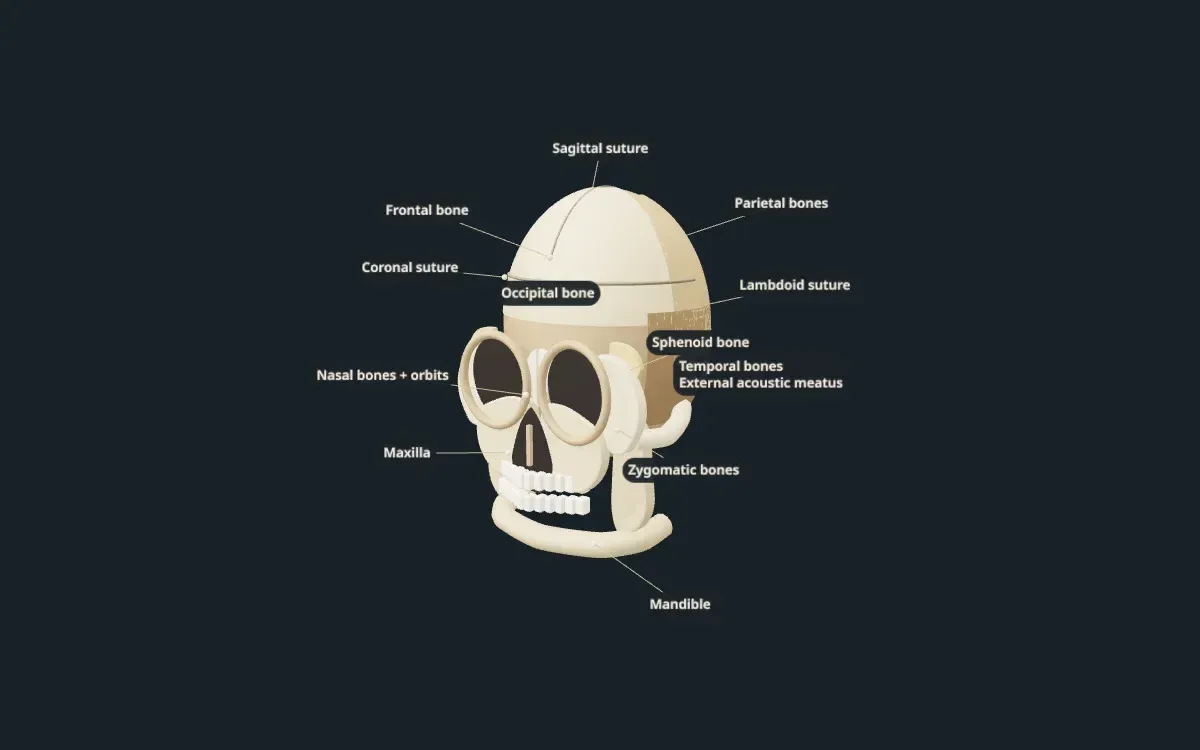
An illustrative teaching model built with Scholarly. Shapes, sizes, and colours are simplified so each part is easy to see.
Free to start. Build one from your own notes, slides, or textbook PDF.
Labelled parts
Bones and sutures of the skull
The 13 parts labelled on the model, and what each one does.
- Frontal bone
- Forms the forehead and the top of the eye sockets.
- Parietal bones
- A pair of bones that form the top and upper sides of the skull.
- Temporal bones
- A pair of bones on the lower sides of the skull. Each holds the structures of the ear.
- External acoustic meatus
- The opening of the ear canal in the temporal bone.
- Occipital bone
- Forms the back and base of the skull. The spinal cord passes through a large opening in it.
- Sphenoid bone
- A butterfly-shaped bone at the base of the skull that links the cranial and facial bones.
- Coronal suture
- The joint between the frontal bone and the two parietal bones.
- Sagittal suture
- The joint down the middle of the top of the skull, between the two parietal bones.
- Lambdoid suture
- The joint between the parietal bones and the occipital bone at the back.
- Nasal bones and orbits
- The nasal bones form the bridge of the nose. The orbits are the eye sockets.
- Maxilla
- The upper jaw. It holds the upper teeth and forms part of the eye sockets and the roof of the mouth.
- Zygomatic bones
- The cheekbones, which also form the outer edge of each eye socket.
- Mandible
- The lower jaw. It is the only bone of the skull that moves.
For class and projects
Using this model for class or a project
For studying, rotate the model all the way around. The frontal bone and face are at the front, the parietal bones across the top, the temporal bones on the sides, and the occipital bone at the back. Then find the three sutures that join them.
For a skull model project or an art reference, use it to check which bone sits where before you sculpt or draw. The colours make the borders between bones easy to follow.
Teachers can show the model on a classroom screen in anatomy, biology, or forensic science lessons. For a course that needs the ethmoid bone, foramina, or the bones of the inner ear, you can build your own version from your notes.
Make your own
Need a model of your own lesson?
This model was built in Scholarly from a short description. You can build one from your own material, labelled in the words your course uses.
- 01
Add your material or describe it
Pick notes, slides, a textbook PDF, or a lecture recording, or type what the model should show.
- 02
Scholarly builds the model
In a few minutes you get an interactive 3D model with labels. A full build uses 1 AI Creation Credit, and you confirm it first.
- 03
Refine it, then use it
Ask for small changes in chat for free. On a paid plan, export a 4K picture, a turntable video, or a .glb file for slides.
FAQ
Skull model questions
What bones make up the skull?
The cranium is made of the frontal, two parietal, two temporal, occipital, sphenoid, and ethmoid bones. The face includes the nasal bones, maxilla, zygomatic bones, and mandible, along with several smaller bones.
What are the sutures of the skull?
Sutures are fixed joints between the skull bones. The main ones are the coronal suture at the front, the sagittal suture along the top, and the lambdoid suture at the back.
Is this skull model anatomically exact?
No. It is a simplified teaching model with each bone coloured so it is easy to tell apart. It is not a medical atlas and is not for clinical use.
Can I use this model for a school project?
Yes, you can use it as a reference while you study or build a physical model. Exporting pictures or video of models you build yourself needs a paid plan.
Can I make my own 3D skull model?
Yes. Sign up, choose 3D model, and describe what you need or add your notes. Scholarly builds a labelled, interactive model in a few minutes. A full build uses 1 AI Creation Credit, and you confirm it first.
Keep exploring
More interactive 3D models
Animal cell 3D model
A labelled cutaway with the nucleus, mitochondria, ER, and Golgi.
Plant cell 3D model
Cell wall, chloroplasts, and the large central vacuole, labelled.
Human heart 3D model
Four chambers, four valves, and the great vessels in a cutaway.
Human brain 3D model
The four lobes, cerebellum, and brainstem, labelled.
Human skeleton 3D model
The major bones of the body, labelled head to toe.
Human eye 3D model
Cornea, lens, retina, and optic nerve in a cutaway.
DNA 3D model
The double helix with its backbone and base pairs.
Atom 3D model
A carbon atom's nucleus and electron shells, Bohr style.
Solar system 3D model
The Sun and eight planets on their orbits.
3D models in Scholarly
How 3D models work, what they cost, and what they can't do yet.
Turn your notes into a 3D model
Build a labelled, interactive model of whatever you are studying or teaching, then make flashcards and a quiz from the same notes.
3D models are in Beta. A full build uses 1 AI Creation Credit; small chat edits are free.