3D Eye Model
Turn this labelled eye around to follow light from the front of the eye to the back. The cutaway shows the layers of the eyeball wall, the lens and the structures that hold it, and where the optic nerve leaves for the brain.
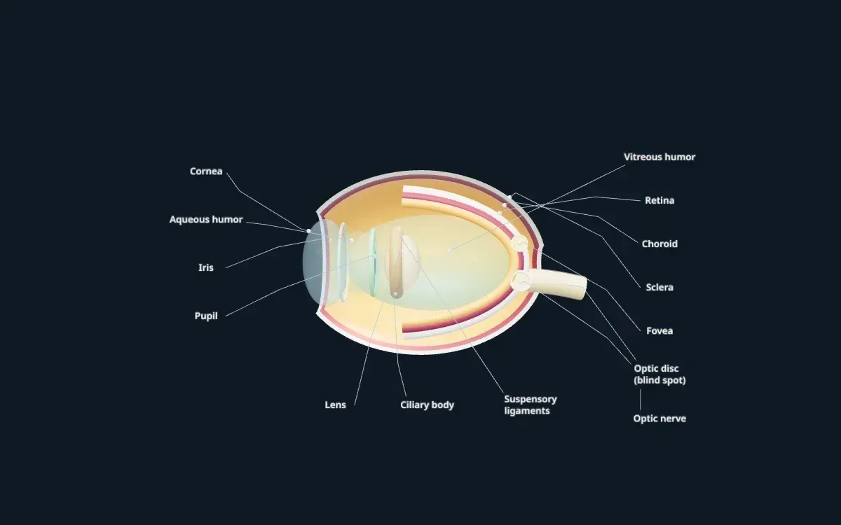
An illustrative teaching model built with Scholarly. Shapes, sizes, and colours are simplified so each part is easy to see.
Free to start. Build one from your own notes, slides, or textbook PDF.
Labelled parts
Parts of the eye
The 13 parts labelled on the model, and what each one does.
- Cornea
- The clear dome at the front of the eye. It does most of the work of bending light into the eye.
- Aqueous humour
- The watery fluid between the cornea and the lens. It nourishes them and keeps the front of the eye in shape.
- Iris
- The coloured ring of muscle that changes the size of the pupil to control how much light gets in.
- Pupil
- The opening in the centre of the iris. It looks black because light passes through it into the eye.
- Lens
- A clear, flexible structure behind the iris that changes shape to focus light on the retina.
- Ciliary body
- A ring of muscle that changes the shape of the lens, and makes the aqueous humour.
- Suspensory ligaments
- Fine fibres that hold the lens in place and connect it to the ciliary body.
- Vitreous humour
- The clear gel that fills the large space behind the lens and keeps the eyeball round.
- Retina
- The light-sensitive layer at the back of the eye. Its rods and cones turn light into nerve signals.
- Fovea
- A small pit in the retina packed with cones. It gives the sharpest, most detailed vision.
- Choroid
- A layer full of blood vessels between the retina and the sclera that supplies the retina.
- Sclera
- The tough white outer layer that protects the eye.
- Optic disc and optic nerve
- The optic nerve carries signals from the retina to the brain. The optic disc, where it leaves the eye, has no light receptors, so it is called the blind spot.
For class and projects
Using this model for class or a project
For studying, follow the path of light on the model: cornea, aqueous humour, pupil, lens, vitreous humour, retina. Then follow the signal from the retina through the optic nerve. Most exam questions about the eye follow one of those two paths.
For an eye model project, use it as a reference for the layers of the eyeball wall: sclera on the outside, then the choroid, then the retina. Keep the lens behind the iris, not in front of it.
Teachers can show the model on a classroom screen while explaining focusing, near and far vision, or the blind spot. For a course that needs the extraocular muscles or the rods and cones up close, you can build your own version from your notes.
Make your own
Need a model of your own lesson?
This model was built in Scholarly from a short description. You can build one from your own material, labelled in the words your course uses.
- 01
Add your material or describe it
Pick notes, slides, a textbook PDF, or a lecture recording, or type what the model should show.
- 02
Scholarly builds the model
In a few minutes you get an interactive 3D model with labels. A full build uses 1 AI Creation Credit, and you confirm it first.
- 03
Refine it, then use it
Ask for small changes in chat for free. On a paid plan, export a 4K picture, a turntable video, or a .glb file for slides.
FAQ
Eye model questions
What are the main parts of the eye?
The cornea, aqueous humour, iris, pupil, lens, ciliary body and suspensory ligaments, vitreous humour, retina and fovea, choroid, sclera, and the optic nerve, which leaves the eye at the optic disc.
How does the eye focus light?
The cornea bends incoming light, and the lens fine-tunes it. The ciliary body changes the lens's shape: it becomes rounder for close objects and flatter for distant ones, so the image lands sharply on the retina.
Is this eye model anatomically exact?
No. It is a simplified teaching model with the parts enlarged and spaced out so each one is easy to see. It is not a medical atlas and is not for clinical use.
Can I use this model for a school project?
Yes, you can use it as a reference while you study or build a physical model. Exporting pictures or video of models you build yourself needs a paid plan.
Can I make my own 3D eye model?
Yes. Sign up, choose 3D model, and describe what you need or add your notes. Scholarly builds a labelled, interactive model in a few minutes. A full build uses 1 AI Creation Credit, and you confirm it first.
Keep exploring
More interactive 3D models
Animal cell 3D model
A labelled cutaway with the nucleus, mitochondria, ER, and Golgi.
Plant cell 3D model
Cell wall, chloroplasts, and the large central vacuole, labelled.
Human heart 3D model
Four chambers, four valves, and the great vessels in a cutaway.
Human brain 3D model
The four lobes, cerebellum, and brainstem, labelled.
Human skull 3D model
The cranial and facial bones and the main sutures.
Human skeleton 3D model
The major bones of the body, labelled head to toe.
DNA 3D model
The double helix with its backbone and base pairs.
Atom 3D model
A carbon atom's nucleus and electron shells, Bohr style.
Solar system 3D model
The Sun and eight planets on their orbits.
3D models in Scholarly
How 3D models work, what they cost, and what they can't do yet.
Turn your notes into a 3D model
Build a labelled, interactive model of whatever you are studying or teaching, then make flashcards and a quiz from the same notes.
3D models are in Beta. A full build uses 1 AI Creation Credit; small chat edits are free.